Dental and maxillofacial
From a single implant to a whole jaw.
Tumour, deformity, trauma or missing teeth: the plan starts with the patient's own scan and your decisions, and reaches theatre as a guide, splint or model made for that patient alone.
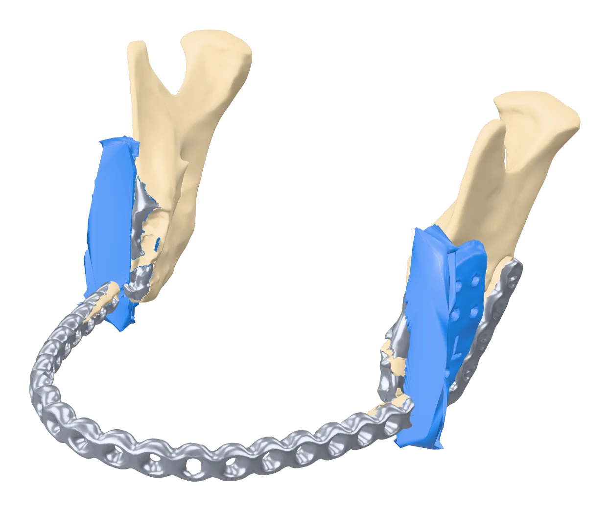
Jaw surgery
Tumour reconstruction, orthognathic and trauma
Ameloblastoma resected and rebuilt with a fibula free flap, jaw correction held by printed splints, fractures rehearsed on true-scale models.
Walk through a real case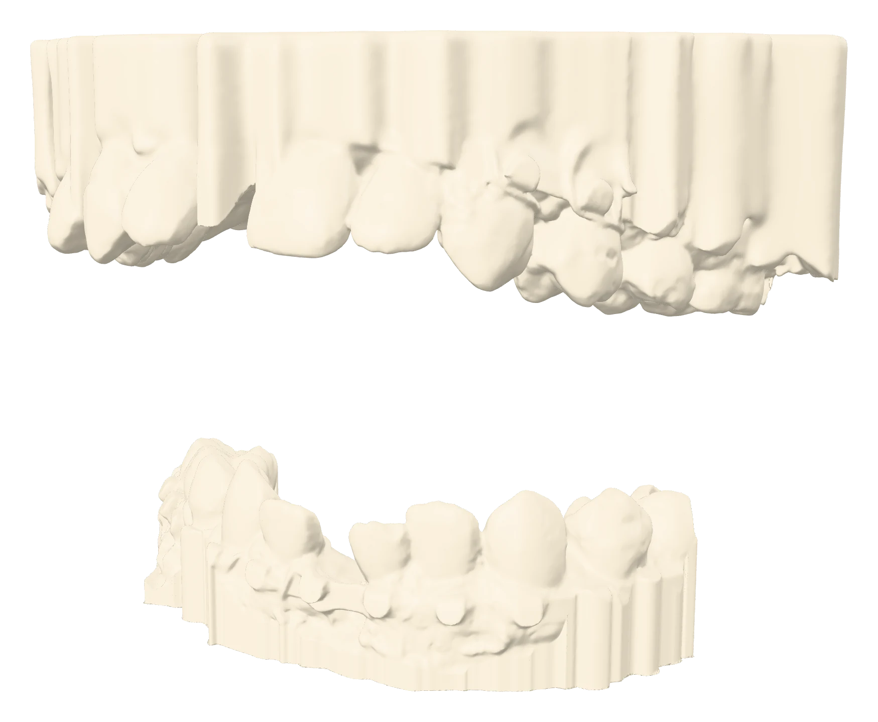
Implants and guides
From intraoral scan to drilling guide
Implant positions planned around the final teeth, then printed as a guide that fits one mouth only.
Implants and guidesFrom an intraoral scan
Upper and lower arches, exactly as scanned.
Crowding, rotations and the gaps a prosthodontist plans around. Add a CBCT and this scan becomes an implant plan and a drilling guide.
Clinical case, 2026. Shown with identifying details removed.
Made in-house, in Nairobi
Everything a dental or jaw case needs, under one roof.
Guides and splints
Implant drilling guides, resection and cutting guides, orthognathic splints: medical-grade, biocompatible and autoclavable.
Dental models
Diagnostic, study and working models from any intraoral scanner, labelled and ready for the lab bench.
Colour anatomical models
Whole skulls and jaws in several colours, pre-operative beside planned, for consent, MDT review and teaching.
Not sure what your case needs? Tell us the clinical question and we will say what we would make.
Evidence
Measured elsewhere. Needed more here.
shorter fibula free flap mandible reconstruction with virtual planning.
Barr 2020, Ann Plast Surg, meta-analysismean deviation at the entry point for implants placed with printed guides, across 2,238 implants.
Tahmaseb 2018, Clin Oral Implants ResOrthognathic surgery planned virtually gave more symmetrical results than traditional planning.
Chen 2021, J Oral Maxillofac SurgRead these numbers as a floor
These studies come from centres that already have navigation, intraoperative imaging and subspecialty teams. Where those are scarce, the printed guide and model are often the only three-dimensional information in theatre, so the gap they close is wider.
Send us the scan.
CT or CBCT as DICOM, intraoral scans as STL. No patient names, please.