Jaw surgery · Live at Kenyatta National Hospital
Every cut decided before the first incision.
Tumour resection and reconstruction, orthognathic correction and facial trauma. One planning service, carried into theatre by guides made for one patient.
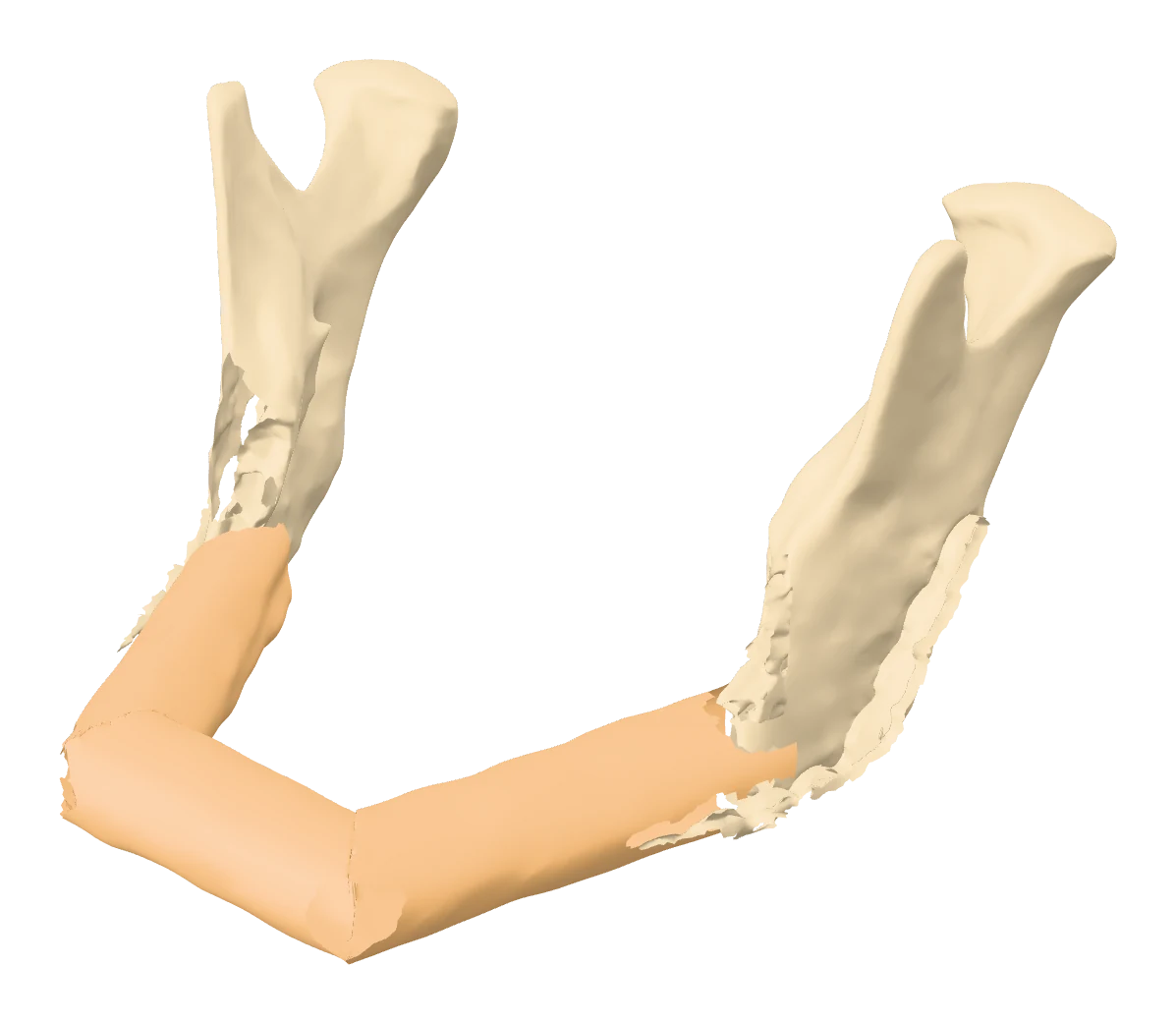
Tumour
Resect and rebuild
Ameloblastoma and other jaw tumours: margins, fibula free flap and dental rehabilitation in one plan.
See the caseDeformity
Orthognathic surgery
Class II, Class III and asymmetry: the jaws moved on screen, then held by printed splints.
The workflowTrauma
Facial fractures
Mandible, midface and orbit at true scale: plates bent before the list, reduction rehearsed.
Fracture planningTumour · A real case, step by step
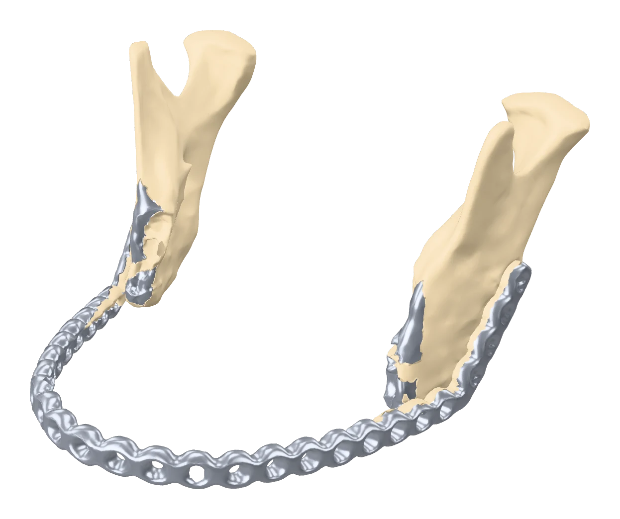
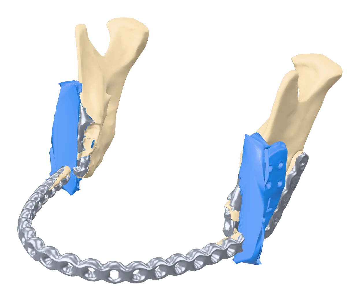
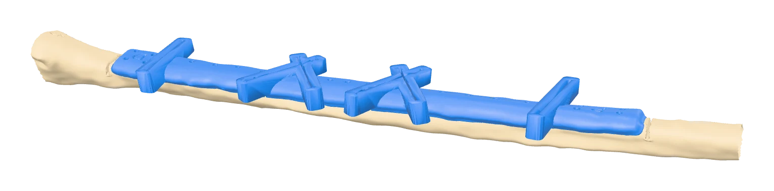
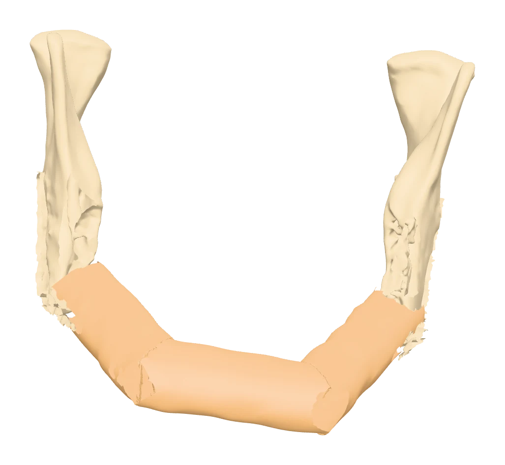
Ameloblastoma: from a plated jaw to a three-segment fibula reconstruction.
Ameloblastoma, Kenyatta National Hospital, 2026. Shown with identifying details removed.
The whole plan at a glance
Resection, harvest and reconstruction, agreed before theatre.
Plan for the teeth now
A fibula seated at the lower border restores the contour but can leave the bone far below the bite. We set its height with you and the prosthodontist in the same session, so an implant-retained prosthesis stays possible. Implants and guides
Part of our oncology work
Jaw tumours are one of several tumour sites we plan: the same approach to margins and reconstruction applies to cardiac and bone tumours. Oncology
Orthognathic surgery
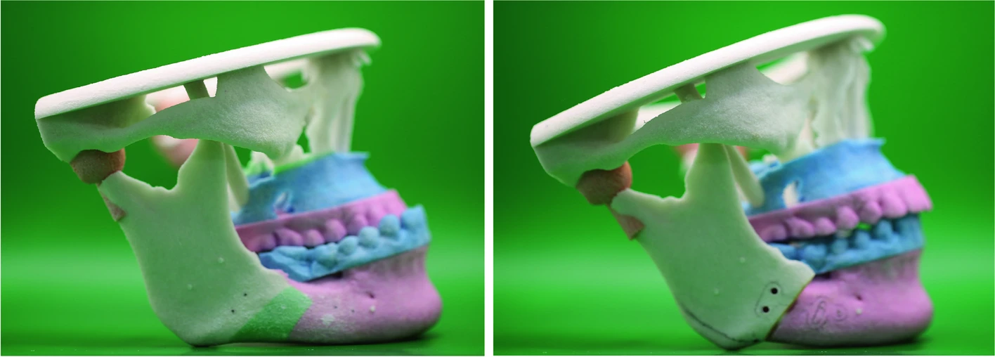
Move the jaws on screen first.
The maxilla and mandible are repositioned in 3D, checked against the bite, the midline and the face, and the plan reaches theatre as printed splints and guides.

What you receive
| Plan | Le Fort I, sagittal split and genioplasty movements, set with the surgeon and orthodontist |
|---|---|
| Splints | Intermediate and final occlusal splints in biocompatible resin |
| Guides | Osteotomy and positioning guides where you want them |
| Models | Colour models before and after, for consent and the team |
| Check | Post-operative scan overlaid on the plan, so you see your own accuracy |
Facial trauma
Bend the plate before the patient is on the table.
Comminuted mandibular, midface and orbital fractures at true scale. For orbital floor and wall repair, the uninjured side is mirrored so the implant is shaped to the anatomy the patient had. Reduction is rehearsed on the model, and the plate is contoured before the list.
Evidence
Shorter operations. More accurate jaws.
shorter fibula free flap mandible reconstruction with virtual planning, with no increase in complications.
Barr 2020, Ann Plast Surg, meta-analysisgonial angle error, planned against free-hand reconstruction.
Pucci 2020, Int J Oral Maxillofac Surg, meta-analysisOrthognathic surgery planned virtually gave more symmetrical results and a more accurate anterior maxilla.
Chen 2021, J Oral Maxillofac Surg; 199 patientsMeasured in centres that already plan in 3D routinely. Where the plan and the guide are the only 3D information in theatre, the gap they close is wider.
Have a jaw to plan?
Send the clinical question. We reply with what the plan can answer, the timeline and the cost.